Digital cadavers bring students a deeper understanding of anatomy, physiology

The challenge of learning human anatomy and physiology can be daunting for many undergraduates: identifying all 206 bones and their every bump, notch and groove; the 320 muscle pairs; the structures and associated tissues of all of the body’s other organ systems — and articulating how all those parts and systems miraculously work together.
To help students master this material, Arizona State University's College of Integrative Sciences and Arts is integrating an innovative visualization tool, the Anatomage Table, into the anatomy and physiology learning experience at the Downtown Phoenix campus.
Adopted by many of the world's leading medical schools, the table is one of only three in Arizona and is the only one to be used in an undergraduate program (the others are used in a graduate program at Northern Arizona University and at Mayo Clinic School of Medicine).
“About 2,000 undergraduate students will engage with the technology in our beginning and advanced anatomy and physiology courses over the course of a year,” said principal lecturer Richard Bauer, who has headed up the faculty of Science, Math and Social Science in the College of Integrative Sciences and Arts since the university opened its Downtown Phoenix campus in 2006.
Bauer’s faculty serves students in all of ASU’s Downtown Phoenix campus colleges, providing foundational and general-studies courses needed by majors in the College of Nursing and Health Innovation, the College of Health Solutions, the Walter Cronkite School of Journalism and Mass Communication, the College of Public Service and Community Solutions, and the College of Integrative Sciences and Arts.
“The majority of the students who take these courses want to enter health-related professions, and this tool will help to more fully prepare them, nicely complementing the anatomical models, texts, plastinatesDonated tissue in which polymers have replaced body fluids. and cadavers,” Bauer said.
RELATED: Anatomage Table is just one of many teaching innovations at CISA
Like a textbook come to life
A month into the spring semester, as the Anatomage technology has begun to be integrated into lab work, the faculty, teaching staff, and students are excited about the table’s features and capabilities.
“The clarity of the images on the table screen is super, like that of a textbook but completely 3-D,” said senior Jessica Lehman, a nursing major who is in her second year working as an instructional assistant in the 200-level anatomy and physiology lab sections. “As a user you have full control of where you move. You can click through each layer of the body, removing one system at a time, or look at an entire system at once. You can choose to searchGet a feel for the experience of using the Anatomage visuals at http://anatomagetable.com. by organ, by body system or by name of a structure.”
Having this additional touchpoint helps students understand context and establish the content in long-term memory, noted lecturer Jeff Kingsbury.
“We all learn in different ways,” Kingsbury said. “And being able to rotate the body from any angle to see all perspectives, students discover for themselves, and can better visualize, the connectedness of body systems and how the function of a structure dictates the anatomical form.
“In thinking about the job of the lungs,” he continued, “with the table we can zoom in to illustrate how the composition of lung tissue enlarges the surface area to impact the whole system.”
Video by Deanna Dent/ASU Now
Sustainability, access and repetition are other benefits of using the table.
For Kingsbury’s advanced dissection students, the table provides some rehearsal context and a great in-between medium between plastinates and cadavers.
“The very first day that we were going to be making cuts we opened up a body on the Anatomage Table and we were able to practice the envelope-shaped cuts we’d be making to open up the rib cage,” said sophomore Kacey Cavanagh, a nursing major. "We were all very nervous about cutting into an actual body, so that was very helpful to see."
The table also helps those unable to do the dissections.
“Some students, because of religious or cultural reasons or past trauma, can’t work with cadavers," noted Bauer, "and so the table also accommodates their learning.
“Physical cadavers have their limits,” he continued. “You can only slice into a body once. Students can slice, if you will, into the digital cadavers a gazillion times. Cadavers can be very messy, and they deteriorate over time. Because of preservation and cleaning techniques it can actually be easier to see some anatomical features in 3-D in the digital environment.”
Junior Kelly Schock has seen this phenomenon firsthand as an instructional assistant for the BIO 494 advanced practicum in human dissection.
Part of her role involves guiding dissection students “to make sure the prosections for BIO 201 and 202 students to observe are meticulously presented and labeled, so that beginning students have access to a near-perfect specimen when viewing a structure for the very first time,” she said.
“The way that fat forms over muscles and organs can hide some structures. Veins, arteries and the lymphatic system are empty and flatten out, and other superficial structures may not present well in a cadaver or may inadvertently ‘go missing’ during the removal of material,” Schock explained. “The cadavers are soaked in a solution to make sure they’re clean enough to handle. You know how your skin gets when you soak in the hot tub for a while? It’s not in its natural state.”
-
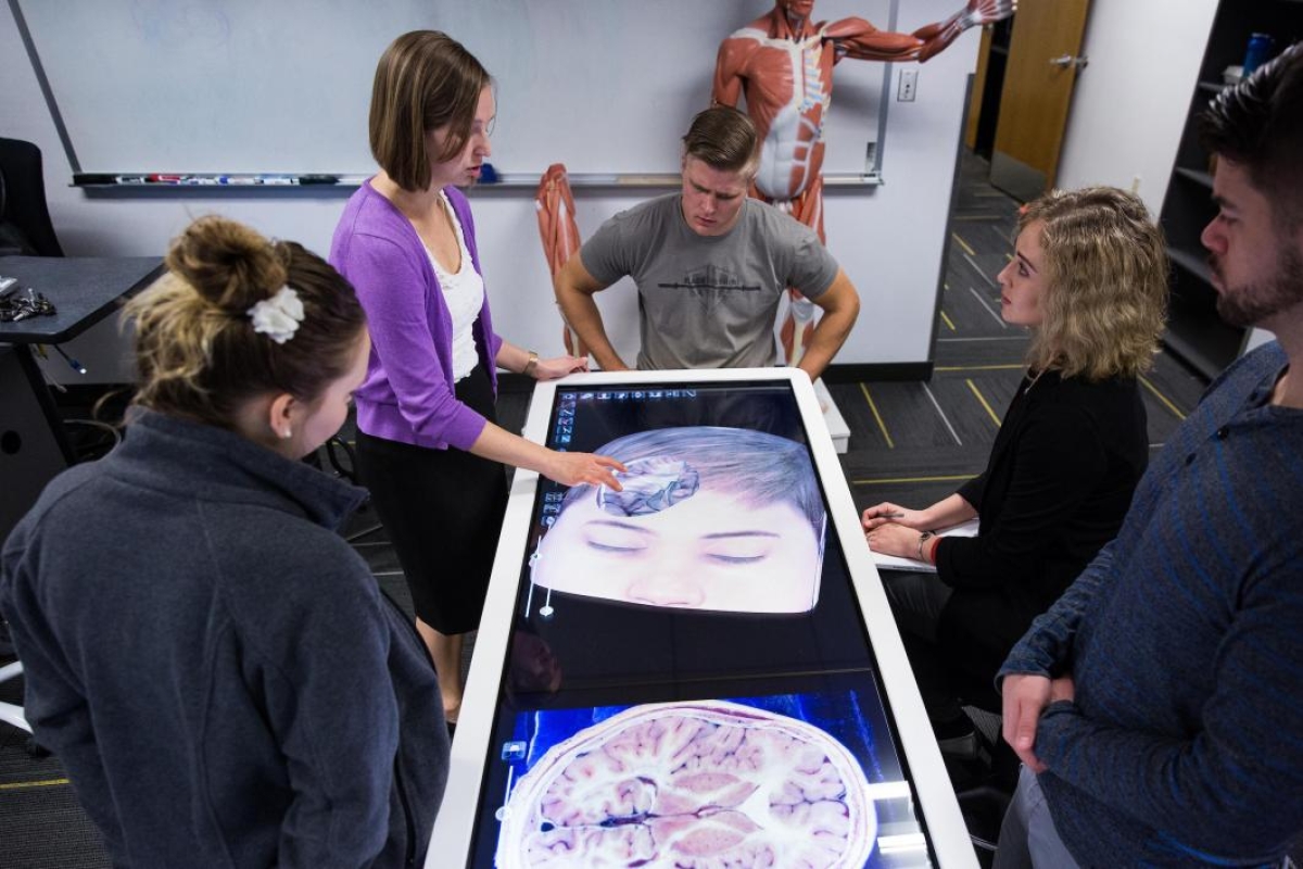
Trainer Samantha Chester (left) walks instructors through the different applications of the Anatomage Table during a training Dec. 8 at the Downtown Phoenix campus. The instructional assistants, who are current undergraduate students, will use the table to help teach labs.
Photo by Deanna Dent/ASU Now
-
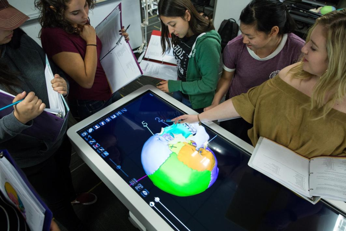
(From left) Team members Vanessa Perez, Evelyn Gutierrez, Ennery Bravo, Cindy Szeto and Taylor Howe test their knowledge of skull bones on the Anatomage Table in their Human Anatomy/Physiology I Lab at the Downtown Phoenix campus on Feb. 7.
Photo by Deanna Dent/ASU Now
-
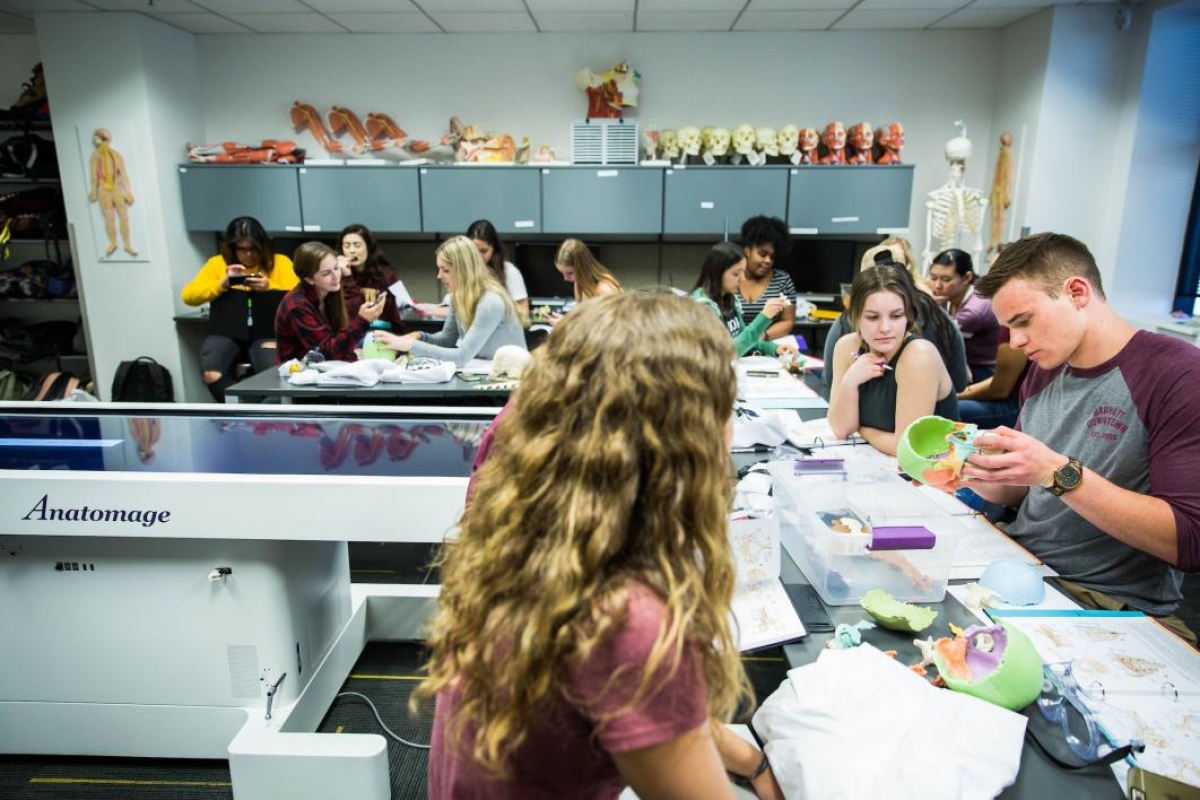
Students practice putting together a 3-D model skull before being tested on their knowledge on the Anatomage Table in their Human Anatomy/Physiology I Lab at the Downtown Phoenix campus on Feb. 7.
Photo by Deanna Dent/ASU Now
-
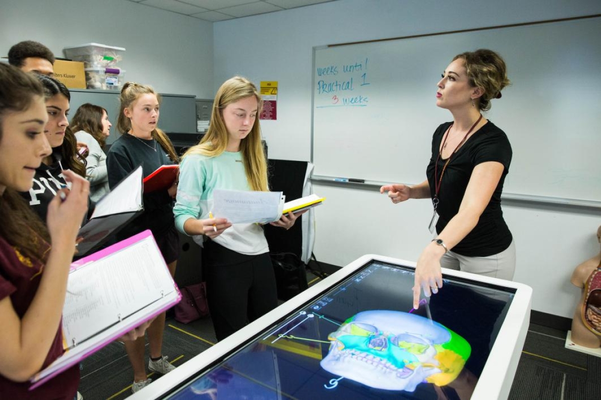
Lab coordinator Jennifer Legere tests her students on their knowledge of human skull bones on the Anatomage Table on Feb. 7.
Photo by Deanna Dent/ASU Now
-
Trainer Samantha Chester (left) walks instructors through the different applications of the Anatomage Table during a training Dec. 8 at the Downtown Phoenix campus. The instructional assistants, who are current undergraduate students, will use the table to help teach labs.
Photo by Deanna Dent/ASU Now
-
(From left) Team members Vanessa Perez, Evelyn Gutierrez, Ennery Bravo, Cindy Szeto and Taylor Howe test their knowledge of skull bones on the Anatomage Table in their Human Anatomy/Physiology I Lab at the Downtown Phoenix campus on Feb. 7.
Photo by Deanna Dent/ASU Now
-
Students practice putting together a 3-D model skull before being tested on their knowledge on the Anatomage Table in their Human Anatomy/Physiology I Lab at the Downtown Phoenix campus on Feb. 7.
Photo by Deanna Dent/ASU Now
-
Lab coordinator Jennifer Legere tests her students on their knowledge of human skull bones on the Anatomage Table on Feb. 7.
Photo by Deanna Dent/ASU Now
More than anything, though, Schock said she has seen how this technology has sparked student interest.
“Linking science and technology this way makes the material exciting for students,” she said. “Now when I throw my lab students bone models to work with, they’ll ask if they can use the table instead. Any science nerd really appreciates it.”
An operating table-sized tablet
The Anatomage technology first came to the attention of Jennifer Legere, anatomy and physiology laboratory coordinator, a few years ago, when she was researching potential teaching tools that would give students more hands-on time.
“We’d been using some 3-D visualization products that were instructor-controlled. I’d be up at the front of the classroom doing the manipulation on two touchscreens with students observing the projection wearing 3-D glasses,” Legere said. “For students, there’s not so much ‘ooh’ and ‘aah’ in that — it’s more like watching something on TV.
“The Anatomage table is very intuitive for our students, who are used to clicking and swiping their phones, tablets and iPads to resize images or go deeper into content. They have picked it up very quickly,” she said. “Manipulating the technology themselves, they’re immediately engrossed in learning and it’s impossible to be detached.
“There was really nothing else out there that compared to this technology,” Legere continued. “With the table we have perpetual access to digital anatomy of three full-size human cadavers, with a range of variation, anomalies and pathology. We also have radiology assets like CT scans and MRIs, and get regular software updates as new content becomes available.
“We can go in and color-code the anatomical components of the Anatomage virtual cadavers to match the color of structures on the magnetic models in our labs,” she added, “so information transfers across platforms seamlessly to our students.”
Legere pitched the product to Bauer in 2014, and the college was able to make the purchase and take delivery in December 2017.
“New content and features were added in the intervening years that made the wait worthwhile,” she noted, “including the capability to switch the table from a horizontal to a vertical orientation, which is helpful when you want to get more students around the table at one time.”
A unique teaching tool
At ASU’s recent Open Door event at the Downtown Phoenix campus on Feb. 2, Lehman and fellow instructional assistant Genesis Rivera, also a senior nursing major, demonstrated the Anatomage technology to community visitors of all ages as Legere buzzed in and out between other demonstrations.
“Could I see the eyes?” asked one child, as Lehman and Rivera encouraged those gathered to pick a body system or organ they’d like to see up close.
“How about the heart?” “The lymphatic system?” “The appendix?” suggested guests. One family group wondered if they could see an image that would help them understand the physiology of a loved one’s recent stroke.
Though the two had only been using the table for a few weeks, their grasp of the material and confidence was unmistakable as visitors peppered them with questions.
Working with the Anatomage technology and being impressed with the company, Schock went to their website two weeks ago and checked out their career postings.
“I emailed their general info address, explaining my background and asked if they ever hired interns,” she said. “They put me in touch with someone, I shared with them my experience and why I’d be interested in working with the company, and they offered me a paid internship. I’ll be working there all summer in Silicon Valley.
“It’s a good reminder that not every path has already been made.
“They want me to help develop curriculum and the questions that go along with the table’s supporting material. I’ll be creating content and preparing data for upcoming software releases, reviewing anatomy accuracy and assisting in corrections and, of course, helping anyone out as needed.
“I came into college thinking I wanted to be a doctor, at one time considered switching to business, but then I took anatomy and said, ‘This is where it’s at!’ ” she said. “I love anatomy and I like people and social interaction, which is why I’m really excited about doing the internship. It’s a perfect combination of the three.”
Top photo: Trainer Samantha Chester walks instructional assistants through the different applications of the Anatomage Table during a training at the Downtown Phoenix campus on Dec. 8. Photo by Deanna Dent/ASU Now
More Science and technology

Hack like you 'meme' it
What do pepperoni pizza, cat memes and an online dojo have in common?It turns out, these are all essential elements of a great cybersecurity hacking competition.And experts at Arizona State…

ASU professor breeds new tomato variety, the 'Desert Dew'
In an era defined by climate volatility and resource scarcity, researchers are developing crops that can survive — and thrive — under pressure.One such innovation is the newly released tomato variety…

Science meets play: ASU researcher makes developmental science hands-on for families
On a Friday morning at the Edna Vihel Arts Center in Tempe, toddlers dip paint brushes into bright colors, decorating paper fish. Nearby, children chase bubbles and move to music, while…