ASU team turns smartphone into a powerful microscope in the fight against infectious diseases
Biodesign researchers shrinks cost of device to diagnose tuberculosis in the field
With smartphones millions of times more powerful than the NASA Apollo computers that sent us to the moon in the 1960s, scientists have been eager to adapt them back here on Earth to better the planet.
That’s exactly what ASU Biodesign Institute researcher Tony Hu and postdoctoral researcher Dali Sun have recently demonstrated in the fight against infectious diseases.
They’ve developed a simple mobile technology for clinics, hospitals and health organizations that are on the front lines of triaging outbreaks around the world.
The innovation takes a $60,000 state-of-the-art technology, called dark-field microscopy, and shrinks the cost down to $2,000 in the hopes of making health-care diagnostics more affordable to limited-resource areas, particularly in the developing world.
Like watching a movie with the lights turned low, dark-field microcopy is a powerful tool for scientists to see more clearly brightly lit samples against a black background, allowing for better contrast.
They hope turning smartphones into handheld microscopes will make an impact as a versatile and powerful new tool in the worldwide fight against infectious diseases, still the world’s No. 1 killer, particularly in the developing world.
“With more and more powerful smartphones equipped with better cameras, this has spurred technology development for now using mobile phone cameras for many medical applications,” said Hu, an associate professor at the Biodesign Institute at Arizona State University’s Virginia G. Piper Center for Personalized Diagnostics and at ASU’s School of Biological and Health Systems Engineering.
“We wanted to develop something that was fast and simple so that potentially anyone can rapidly diagnose an infectious disease.”
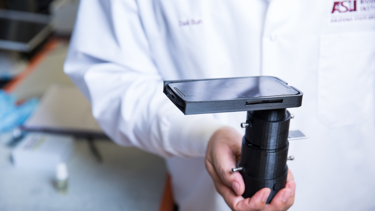
Using 3-D printing, Sun custom fabricated their first prototype, which contains an easy-to-use mobile phone attachment that slides on like a smartphone case, and a condenser to help focus light onto a sample.
Video by Deanna Dent/ASU Now
Ironically, the toughest fit turned out to be finding the right light source. After scouring online, Sun eventually found a $1 battery-operated LED light that was the perfect fit.
The microscope housing also contains a slide reader and specially coated slides that are customized to detect a specific infectious disease, and also assess the severity of infection.
One of their first applications was a novel nanoparticle-based serum diagnostic assay for tuberculosis, a notoriously difficult-to-diagnose infection.
The test is sensitive enough to give a result from just a single drop of liquid that is prepared from a patient’s blood sample using a custom, patented sample prep kit that Hu’s lab has developed.
If TB is present, it can be seen as a red color with the smartphone dark-field microscope.
“If you see a high intensity of red, that means the person is TB positive,” said Sun. “If you don’t see any red, that means they are TB negative.”
“These assays yielded robust results that were similar to, albeit less sensitive than, those obtained with a much more expensive and cumbersome desktop dark-field microscope system,” said Hu.
The device is only the first prototype, with an ultimate aim of making the dark-field microscope attachments less than $100.
And their nanoparticle-based detection system has the potential to be adaptable to a number of other infectious diseases.
And the beauty of their system is that it has the potential to be adaptable to a number of other infectious diseases, to aid in the early diagnosis and evaluation of more effective treatments.
Top photo: Postdoctoral researcher Dali Sun holds a mobile phone adaptation of a dark-field microscope, which could be used in developing areas where access to a traditional desktop dark-field microscope might not be possible. Photo by Deanna Dent/ASU Now
More Health and medicine

First exchange student for Biodesign Institute Europe bridges labs 5,000 miles apart
This spring semester, Grace Colley traveled to Arizona State University and became the first student to participate in the Biodesign Institute Europe student exchange program. In doing so, she helped…
College of Health Solutions hosts visit from leading expert in genomic research
Some fortunate Arizona State University faculty, staff and students were able to gain valuable insights and perspective during a visit by one of the country’s leading figures in health and scientific…

Indigenous ASU research team recommends assistance for tribal members still reeling from COVID-19’s effects
When Matt Ignacio’s tribe, the Tohono O’odham Nation, donated $1 million to Arizona State University to support COVID-19 research, he applied for some of the money to understand and report any…